Diagnostic Imaging
Astera Health offers advanced diagnostic imaging techniques and technologies to Central Minnesota residents. Our expert team performs over 25,000 imaging procedures annually.
Whether you need a medical imaging procedure for an injury, a healthcare screening, or an image-guided procedure, you can be confident in the imaging services you receive at Astera. In addition to providing compassionate care, we’re an American College of Radiology-accredited facility for computed tomography (CT), magnetic resonance imaging (MRI), and ultrasound, and mammography.
A closer look at diagnostic imaging
Diagnostic imaging allows healthcare providers to see internal parts of the body, such as blood vessels, bones, muscles, or organs. Images created by diagnostic imaging technology are used to:
- Diagnose or rule out medical conditions
- Guide treatment decisions
- Monitor how well treatments are working
- Screen for health conditions, including cancer and osteoporosis
A radiologist, a provider with special training in imaging technology, reviews all images. The radiologist may meet with you to go over your results or send their findings to your family medicine provider to review with you. If your tests show abnormalities, your provider will talk with you about your next steps.
Advanced imaging technology
Different kinds of imaging technology are designed to gather specific types of information. Depending on your needs, your provider may recommend one or more imaging tests, including:
- 3D mammography. A computer creates three-dimensional breast images from several 2D X-rays.
- Bone densitometry, or DEXA scan. High- and low-energy ionizing radiation passes through the body to help detect thinning bones.
- Computed tomography, or CT scan. Several detailed X-ray image slices combine to create a 3D image of structures inside the body.
- Echocardiography. Echoes of high-frequency sound waves create moving or still images of the heart.
- Fluoroscopy. High-energy ionizing radiation beams move through the body to create real-time, movie-like images of the inside of the body.
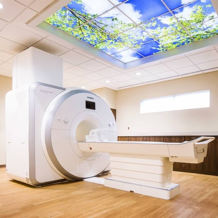
- Magnetic resonance imaging, or MRI. 3D images of internal structures are created using powerful magnetic fields and radio waves.
- Nuclear cardiology study, or stress test. Images of radioactive material injected into the bloodstream show how the heart is working during exercise.
- Positron emission tomography, or PET scan. Diagnostic imaging paired with nuclear medicine shows body function by tracking a radioactive tracer injected into the body.
- Ultrasound. High-frequency sound waves move through the skin and bounce off internal body structures to produce images.
- Videofluoroscopic swallow study. This specialized imaging study is conducted alongside our speech therapy department and uses a continuous X-ray image to create movie-like images.
- X-rays. Ionizing radiation rays pass through the body, producing still images of internal structures.
Now offering PET/CT Scans
Positron Emission Tomography/Computed Tomography (or PET/CT) Imaging combines the functional information from a PET exam with anatomical information from a CT exam in one single exam.
This technology provides new information that can alter a patient’s treatment plan to better target the cancer in approximately one-third of cases. It can also detect additional tumors a single exam can miss.
PET/CT exam results may have a major impact
on your diagnosis, and the course of treatment selected
by your physician.
A PET/CT study not only helps your physician diagnose
a problem, it also helps your physician predict the
likely outcome of various therapeutic alternatives,
pinpoint the best approach to treatment, and monitor
your progress. If you are not responding as well as
expected, you may be switched to an alternative
therapy.
When you arrive, check-in at the Imaging Registration desk. The technologists will review your history and any past exams.
For the PET portion of the exam, you will receive
an injection of radioactive material similar to what
is used for bone scans and other nuclear medicine
exams. PET radiopharmaceuticals
lose their radioactivity very quickly (two hours) and
only very small amounts are injected. In all cases, the
majority of radioactivity will be eliminated from the
body approximately six hours after injection.
After your injection, you will be asked to wait
in our injection suite for one hour while the
radiopharmaceutical is distributed. During this time
you will be asked to relax.
During the exam, you will lie very still on a comfortable
table that will move slowly through the scanner
as it acquires the information needed to generate
diagnostic images.
Plan to spend two to three hours with us, however, the
scan should only last between 15 and 45 minutes.
The exam time can vary depending on what we are
looking for and what we discover along the way.
You may return to the designated area as soon
as the exam is complete. Unless you’ve received
special instructions, you can eat and drink
immediately. Drinking lots of fluids soon after the
exam will help remove any radiopharmaceuticals
that may still be in your system.
Following your exam, we’ll begin preparing the results
for review by our interpreting physician, and then by
your physician, who will tell you what we’ve learned.
Be assured that PET/CT exams are a safe and effective
diagnostic procedure. The radiopharmaceuticals used
in PET do not remain in your system long, so there’s
no reason to avoid interacting with other people
once you’ve left. To be extra safe, wait for a few hours
before getting too close to an infant or anyone pregnant.
Minimally invasive image-guided procedures
In addition to diagnostic imaging, Astera offers several minimally invasive interventional radiology procedures. During these procedures, your radiologist uses real-time imaging technology as a guide. Compared to other surgical techniques, these procedures typically cause less pain and have a faster recovery period.
Our special interventional radiology procedures include:
- Arthrography
- Myelography
- Image-guided back, joint, and neck injections
- Radiofrequency ablation
- Stereotactic breast biopsies and other guided biopsy techniques
Diagnostic imaging and radiation safety
Although some diagnostic imaging tests, such as X-rays and CT scans, expose patients to ionizing radiation and radioactive materials, the benefits of these tests generally outweigh the risks. Imaging technology and techniques are also designed to minimize radiation exposure. Talk with your family medicine provider before your imaging procedure if you’re pregnant or have questions or concerns.
CONTACT US
Appointments:
Do you have an order for diagnostic imaging? Call 218-631-3510 to schedule an appointment.
Hours:
Monday – Friday: 8 a.m. – 5 p.m.
We also offer on-call coverage for emergencies during off hours.
Astera Health is proudly accredited for MRI, Ultrasound, and CT.
Why choose Astera Health?
When you need medical care, you want to ensure you’re getting the best care possible. Astera strives to ensure that all patients always receive a high level of service. Patients love our clinics for all of the following reasons:
Care for all ages
We provide comprehensive services for all ages, from young children to older adults.
Personalized treatment
We develop customized treatment plans to meet the unique needs of every patient, ensuring they receive the best care.
Compassionate care
Our medical staff takes a personal interest in all our patients, providing compassionate care in your time of need.